
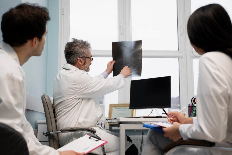
When a health problem causes pain, unusual symptoms, or changes that cannot be identified through a physical examination alone, diagnostic imaging can provide important clues. Radiology in Paschim Vihar includes imaging-based investigations that help doctors examine structures inside the body and make informed decisions about further care. From a simple X-ray to ultrasound, CT scans, and MRI, different imaging techniques serve different purposes. Understanding how they work and when they may be recommended can help patients feel more prepared when their doctor advises an imaging test.
Radiology helps detect health problems early by creating detailed images of organs, bones, tissues, and other internal structures. X-rays, ultrasound, CT scans, and MRI can help doctors identify abnormalities, monitor known conditions, investigate symptoms, and guide treatment decisions when clinically appropriate.
What Is Radiology and Why Is It Important?
Radiology is a medical field that uses imaging technology to examine the inside of the body. These images can help healthcare professionals investigate symptoms, identify abnormalities, assess injuries, and monitor certain conditions over time.
Importantly, not every patient needs the same type of scan. The appropriate examination depends on factors such as symptoms, medical history, age, suspected condition, and the clinical question the doctor needs to answer.
Early investigation can sometimes help identify a problem before it becomes more complicated. However, an imaging test does not automatically mean that a serious disease is present. Results need to be interpreted alongside the patient’s symptoms and clinical examination.
How Radiology in Paschim Vihar Supports Early Detection
Access to appropriate diagnostic imaging can make the evaluation of many health concerns more efficient. Depending on the situation, radiology may help doctors:
- Examine an injury or suspected fracture
- Investigate persistent pain or swelling
- Assess certain abdominal or pelvic symptoms
- Identify changes in organs or soft tissues
- Investigate respiratory symptoms when imaging is clinically indicated
- Monitor an already diagnosed condition
- Evaluate whether further testing may be necessary
The main advantage is that imaging can provide information that cannot always be obtained through a physical examination alone.
X-Ray for Bones and Chest Evaluation
X-rays are among the most commonly used imaging examinations. They use a small amount of ionizing radiation to create images of structures such as bones and the chest.
Doctors may recommend an X-ray when they need to evaluate a suspected fracture, certain joint problems, or particular chest-related concerns. Because X-rays are quick and widely available, they can be useful when a straightforward image can answer the clinical question.
Ultrasound for Real-Time Imaging
Ultrasound uses sound waves rather than ionizing radiation to produce images. It is commonly used to examine areas such as the abdomen, pelvis, thyroid, and certain soft tissues.
It can also be useful in pregnancy care because ultrasound provides real-time images without using ionizing radiation. Depending on the clinical situation, Doppler ultrasound can also assess blood flow through certain blood vessels.
CT Scans for Detailed Cross-Sectional Images
Computed tomography, or CT, combines X-ray images to produce detailed cross-sectional views of the body. It can provide more information than a conventional X-ray for many clinical questions.
Doctors may use CT when they need to assess certain injuries, internal structures, or other conditions that require more detailed imaging. Some CT examinations use contrast material to improve visualization of particular tissues or blood vessels.
Because CT involves ionizing radiation, the test should be performed when its expected clinical benefit justifies the exposure.
MRI for Soft-Tissue Assessment
Magnetic resonance imaging, commonly called MRI, uses a strong magnetic field and radio waves to produce detailed images. It does not use ionizing radiation.
MRI can be particularly useful for evaluating soft tissues, including the brain, spine, muscles, ligaments, and certain organs. Patients with some implanted medical devices or metal fragments may need special assessment before undergoing an MRI.
When Should You Consider Diagnostic Imaging?
Radiology should not be viewed as a routine test that everyone needs. A doctor usually recommends imaging when the patient’s symptoms, examination findings, medical history, or risk factors suggest that an image could provide useful information.
You may be advised to undergo diagnostic imaging when you have:
- Persistent or unexplained symptoms
- A suspected bone or joint injury
- Certain abdominal or pelvic complaints
- Symptoms requiring evaluation of internal organs
- A condition that needs monitoring
- A clinical concern that cannot be adequately assessed through examination alone
If symptoms are new, severe, persistent, or worsening, speak with a qualified healthcare professional rather than choosing an imaging test independently.
What Happens During a Radiology Examination?
The process depends on the imaging technique. Before the examination, the radiology team may ask about your symptoms, medical history, medications, allergies, pregnancy status when relevant, or previous imaging studies.
Some examinations require specific preparation. For example, certain abdominal scans may require fasting, while some contrast-enhanced examinations involve additional instructions.
During the test, you may need to remain still or follow breathing instructions. Some scans take only a few minutes, while more detailed examinations can take longer.
Afterward, a radiologist reviews the images and prepares a report for the referring healthcare professional. The doctor then interprets the findings in the context of your overall health.
Choosing a Radiology Centre: What Should You Check?
If your doctor recommends imaging, choosing an appropriate diagnostic facility matters. When looking for Radiology in Paschim Vihar, consider factors such as:
- Availability of the required imaging test
- Appropriate and well-maintained equipment
- Qualified radiology professionals
- Clear patient preparation instructions
- Proper radiation-safety practices
- Accurate reporting and communication
- Convenient location and appointment availability
You can also ask whether previous reports or images should be brought to the appointment. Comparing new images with earlier studies can sometimes help healthcare professionals assess changes over time.
Why Professional Interpretation Matters
An imaging report should not be interpreted in isolation. A finding that appears on a scan may have different meanings depending on the patient’s symptoms, age, medical history, and other investigations.
Radiologists are trained to interpret medical images and communicate relevant findings in a structured report. Your treating doctor can then explain what those findings mean for your particular situation and whether further evaluation is appropriate.
This approach helps prevent unnecessary anxiety from reading imaging terminology without clinical context.
5 FAQs
What types of imaging are commonly available through radiology services?
Common diagnostic imaging methods include X-rays, ultrasound, CT scans, and MRI. The appropriate test depends on the symptoms and the medical question being investigated. A doctor may recommend one examination over another based on factors such as the body area involved, suspected condition, medical history, and the benefits and risks of the test.
Is radiology useful for detecting health problems early?
Yes, appropriate imaging can help identify abnormalities or changes that may not be apparent from symptoms or physical examination alone. However, imaging is not a universal screening tool for every disease. The value of a scan depends on the patient’s individual circumstances and whether the examination is clinically indicated.
Which is better: CT scan, MRI, or ultrasound?
There is no single imaging test that is best for every condition. Ultrasound can be useful for many soft-tissue and abdominal examinations, CT provides detailed cross-sectional images, and MRI is particularly valuable for many soft-tissue conditions. A healthcare professional selects the examination based on the clinical question.
Should I prepare before a diagnostic imaging test?
Preparation varies according to the examination. Some scans may require fasting, while others need no special preparation. If contrast material is planned, the facility may ask about allergies, medications, kidney problems, or other relevant health information. Always follow the specific preparation instructions provided by your healthcare team.
How do I choose a radiology centre in Paschim Vihar?
Look for a centre that offers the imaging examination you need, follows appropriate safety practices, has qualified professionals, and provides clear reporting and patient instructions. Location and appointment convenience are also useful considerations. Most importantly, choose imaging based on a healthcare professional’s recommendation rather than ordering unnecessary tests yourself.
Conclusion
Radiology plays an important role in modern diagnosis by giving healthcare professionals a clearer view of structures inside the body. Depending on the clinical need, X-rays, ultrasound, CT, or MRI can help investigate symptoms, assess injuries, monitor known conditions, or identify abnormalities that may need further attention. Radiology in Paschim Vihar can therefore be a valuable part of appropriate medical evaluation when recommended by SODHI Nursing Home. The right imaging test depends on the individual patient and the question that needs to be answered. If you have persistent or unexplained symptoms, discuss them with your doctor to determine whether diagnostic imaging or another form of evaluation is appropriate.