
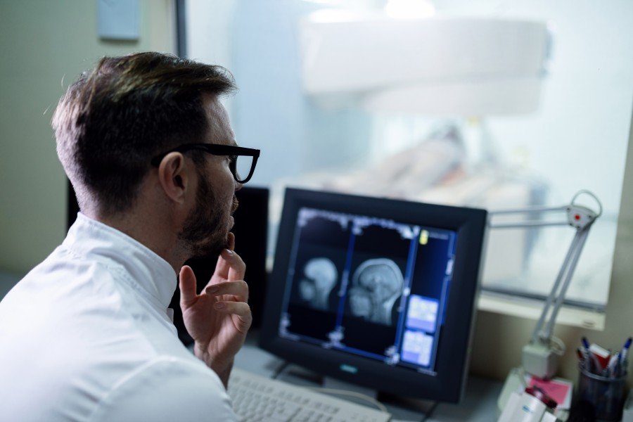
When a doctor needs more information about what is happening inside the body, diagnostic imaging can provide valuable clues. Radiology in Paschim Vihar includes a range of imaging-based investigations that can help healthcare professionals evaluate bones, organs, soft tissues, blood vessels, and other structures.
Different imaging tests serve different purposes. An X-ray may be useful for examining bones, while ultrasound can provide real-time images of certain organs and tissues. CT and MRI scans offer more detailed views and may be recommended when additional information is needed.
Radiology in Paschim Vihar involves diagnostic imaging tests such as X-rays, ultrasound, CT scans, and MRI. These examinations help doctors evaluate injuries, infections, organ abnormalities, and other health concerns. The appropriate test depends on the symptoms, clinical history, body area being examined, and the information the treating doctor needs.
What Is Radiology and Why Is It Important?
Radiology is a medical field that uses imaging techniques to examine structures inside the body. These images can support diagnosis, treatment planning, monitoring, and follow-up.
Importantly, an imaging test is not usually selected simply because it is available. A doctor considers the patient’s symptoms, medical history, physical examination, and suspected condition before deciding which investigation may be appropriate.
Radiology can therefore play an important role when a physical examination alone does not provide enough information.
Common Types of Diagnostic Imaging Tests
X-Ray
X-rays use a small amount of ionizing radiation to create images of structures inside the body. They are commonly used to examine bones and the chest.
Doctors may recommend an X-ray when evaluating suspected fractures, certain lung or chest problems, joint issues, or other conditions where an X-ray can provide useful information.
Although X-rays are quick, they are not suitable for every diagnostic question. The treating professional determines whether the examination is appropriate.
Ultrasound
Ultrasound uses high-frequency sound waves rather than ionizing radiation to create images.
It is commonly used to examine areas such as the abdomen, pelvis, thyroid, and certain soft tissues. Ultrasound is also widely used during pregnancy for appropriate obstetric assessment.
One advantage is that ultrasound can provide real-time imaging. However, image quality can depend on the body area being examined and other factors, so it may not replace CT or MRI when more detailed imaging is required.
CT Scan
Computed tomography, or CT, combines multiple X-ray images to produce detailed cross-sectional images of the body.
A CT scan can provide more detailed information than a conventional X-ray and may be used to evaluate injuries, internal bleeding, certain infections, abdominal problems, and other medical concerns.
Some CT examinations involve contrast material to make particular structures easier to see. Patients should inform their healthcare team about relevant allergies, kidney problems, pregnancy, or other medical considerations before an examination.
MRI Scan
Magnetic resonance imaging uses a strong magnetic field and radio waves to produce detailed images of the body’s structures. Unlike X-rays and CT scans, MRI does not use ionizing radiation.
MRI can be particularly useful for evaluating soft tissues, the brain, spinal cord, joints, muscles, and ligaments. The examination can take longer than some other imaging tests, and patients with certain implanted medical devices may require additional safety assessment before an MRI.
How Is the Right Imaging Test Selected?
The most appropriate diagnostic scan depends on the clinical question rather than simply choosing the most advanced technology.
A doctor may consider:
- The patient’s symptoms and duration
- The body part that needs evaluation
- Previous medical history
- Results of physical examination
- Earlier laboratory or imaging findings
- Whether radiation exposure should be avoided or limited
- Whether contrast material may be required
- The level of detail needed
For example, a suspected bone fracture may initially be evaluated with an X-ray, while a problem involving soft tissues may require a different imaging approach.
When Should You Discuss Imaging With a Doctor?
Not every symptom requires an imaging test. In many situations, a doctor can determine whether imaging is useful after discussing the symptoms and performing an examination.
Imaging may become relevant when there is a suspected injury, persistent unexplained symptom, concern about an internal organ, or a need to monitor an already identified condition.
Patients should also share relevant information before an examination, particularly pregnancy or possible pregnancy, allergies, kidney problems, previous reactions to contrast material, and implanted medical devices.
Choosing a Radiology Facility in Paschim Vihar
If your doctor recommends diagnostic imaging, choosing an appropriate facility involves more than location.
Consider whether the facility offers the specific examination requested, follows appropriate patient-safety procedures, provides clear preparation instructions, and has qualified professionals involved in image interpretation.
It is also useful to ask whether previous reports or images should be brought to the appointment. Comparing current and earlier studies can sometimes provide important clinical context.
Most importantly, follow the preparation instructions provided for your particular examination. Some scans may require fasting, while others may involve specific instructions regarding clothing, medications, or contrast material.
Why Accurate Reporting Matters
Producing an image is only one part of diagnostic imaging. The images must also be assessed and reported appropriately.
A radiology report describes relevant findings and may help the treating doctor understand what the images show. The final medical interpretation should always be considered alongside symptoms, examination findings, medical history, and other investigations.
Patients should therefore discuss imaging reports with their treating doctor rather than attempting to diagnose a condition from the report alone.
Frequently Asked Questions
What are the most common radiology tests?
Common diagnostic imaging tests include X-rays, ultrasound, CT scans, and MRI. Each has different applications. X-rays are commonly used for bones and chest imaging, ultrasound is useful for many soft-tissue and abdominal examinations, CT provides detailed cross-sectional images, and MRI is particularly useful for many soft-tissue and musculoskeletal evaluations.
When does a doctor recommend an imaging test?
A doctor may recommend imaging when symptoms, physical examination findings, or medical history indicate that additional information is needed. Imaging can help evaluate injuries, persistent symptoms, suspected internal problems, or known conditions requiring monitoring. The choice of test depends on the clinical question and should be determined by an appropriate healthcare professional.
Is ultrasound safer than an X-ray?
Ultrasound does not use ionizing radiation, whereas X-rays do. Ultrasound uses sound waves to produce images and is commonly used for several abdominal, pelvic, and soft-tissue examinations. However, the two tests serve different purposes, so one should not automatically be considered a substitute for the other.
How should I prepare for a radiology test?
Preparation depends on the examination. Some imaging tests require no special preparation, while others may require fasting or specific instructions. If contrast material is planned, the healthcare team may ask about allergies, kidney problems, medications, or previous reactions. Always follow the preparation instructions provided by the imaging facility or doctor.
How do I choose a radiology facility in Paschim Vihar?
Look for a facility that provides the specific examination recommended by your doctor and follows appropriate imaging and patient-safety procedures. Ask about preparation requirements, reporting arrangements, and whether you need to bring previous scans or reports. The most suitable facility depends on the examination required and your individual clinical circumstances.
Conclusion
Radiology can provide important information when a doctor needs to investigate symptoms, evaluate an injury, or understand what is happening inside the body. Radiology in Paschim Vihar may include examinations such as X-rays, ultrasound, CT scans, and MRI, with each test serving a different diagnostic purpose. The right examination depends on the patient’s symptoms, medical history, and the clinical question being investigated. If your doctor recommends diagnostic imaging, follow the preparation instructions carefully and discuss any relevant medical conditions or concerns beforehand. For local patients, choosing SODHI Nursing Home means considering the required test, safety procedures, reporting process, and professional interpretation before proceeding.